The sensation of self
Internal states and visual perception
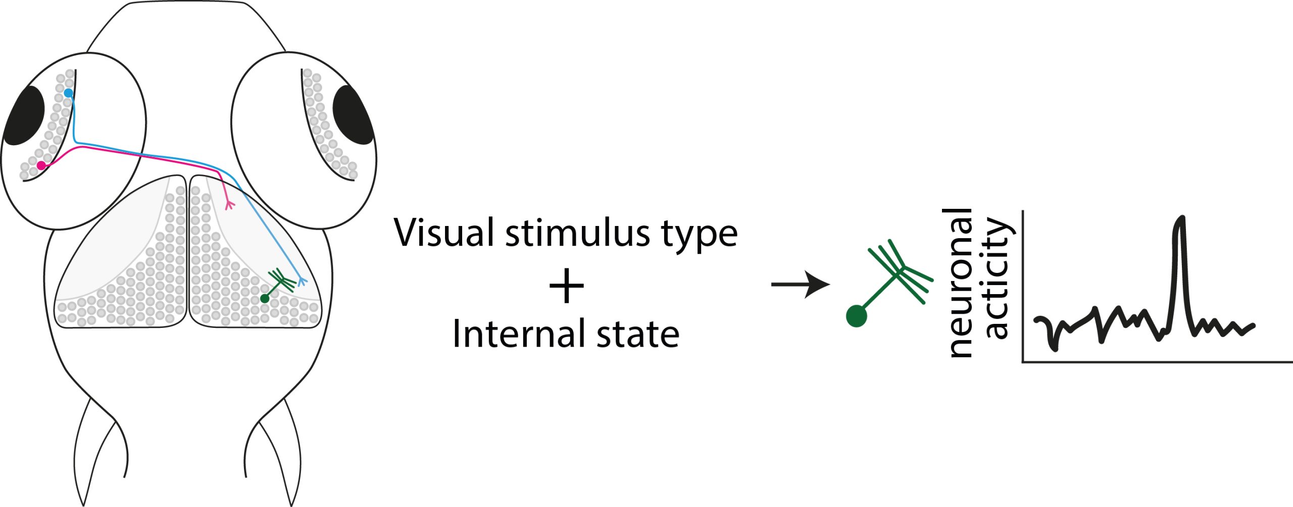
Our brain's response to what we see isn’t fixed - it can change depending on factors like hunger or alertness. For example, neurons in visual areas like the retina and the superior colliculus in mammals can react more or less strongly depending on how awake or hungry an animal is. In zebrafish, hunger can even shift the visual tuning of neurons, such as the preferred size of visual objects.
Interestingly, certain receptors involved in hunger and fullness signals (called melanocortin receptors) are found in the zebrafish optic tectum, suggesting a direct link between these signals and visual processing. While these receptors are known to regulate feeding behavior through other parts in the brain, their role in adjusting visual perception is still a mystery. We are now exploring whether these receptors influence how fish visually perceive prey or predators and how they see and react to their environment.


Transcriptional maps for vision
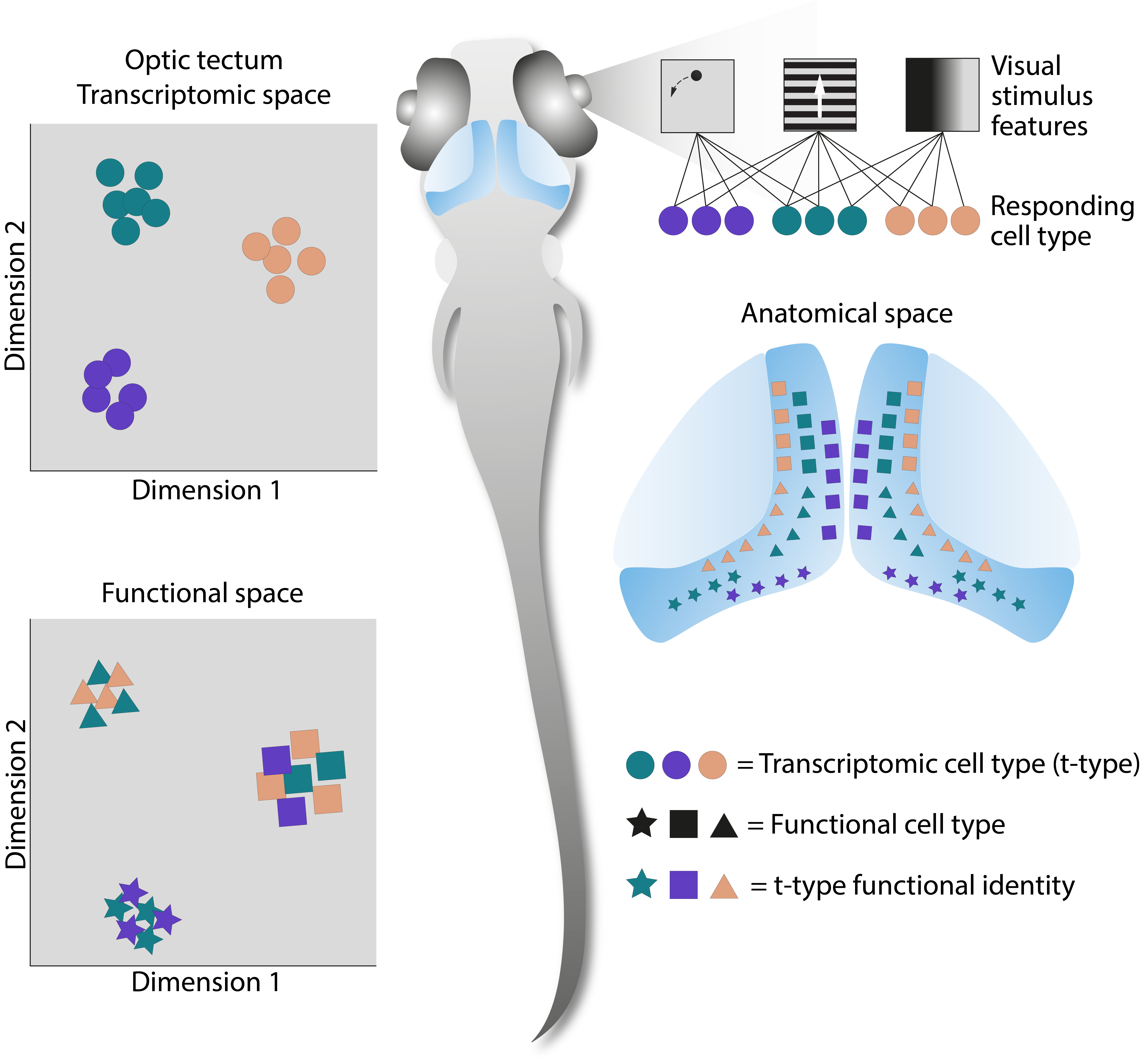
Zebrafish, which hunt prey located above their visual horizon, use specific retinal cells to detect items in the upper part of their vision. These cells send signals to a specific region in the optic tectum that aids in prey capture. In contrast, bottom-feeding fish focus on food below them, which requires a different set of neurons for detection.
In zebrafish, distinct regions of the optic tectum are specialized for processing different visual tasks, but it is unclear whether other fish species have similar setups or if their optic tectum is organized differently to suit their unique behaviors and environments.
To explore this, we compare the visual brain areas and adaptations of different fish species. This will help us understand whether they use the same basic brain wiring in different ways or if their visual systems are entirely restructured to meet their specific needs.